incisive foramen radiograph
The presence of the cyst is presumed if the width of the foramen exceeds 1 cm or if enlargement can be demon-strated on successive radiographs. Type 7 a represents the involvement of the primary hard palate lying anterior to the incisive foramen and posterior to the alveolus b the involvement of.
It m ay be seen as a periapical lesion Fig4C.

. 3B can be seen leading to the incisive fora-. Appears as aV-shaped radio-opaque structure in the midline above the incisive foramen. The radiographic projection angle mental foramen may lead to diagnostic problem.
Here a posterior-occlusal view of a skull demonstrates the incisive foramen. In the human mouth the incisive foramen also known as. It can be single or multiple.
Or the anterior palatine fossa it usually appears as a prominent radiolucent area above or between roots of two central incisors it usually appears round or oval in shape and doesnt exceed 6mm in. As age of the subjects increased incisive foramen diameter and incisive canal length were. The foramen leads to a short canal that connects the nasal and oral cavities.
Although occasionally observed in radiographic examinations of the incisor area of the maxilla nasopalatine duct cysts were. Our goal is to evaluate identification of MIC by both panoramic radiograph PAN and cone-beam computed tomography CBCT. The median suture of the palate see figure 3-23 may appear as a radiolucent line extending posteriorly from the alveolar border.
The mean width of the foramen labiopalatally and mesiodistally was 312 094 mm and 323 098 mm respectively. It transmits the greater palatine artery and. Interpretation of incisive foramen on radiographs.
The incisive foramen is used as an. On periapical x-ray images the incisive foramen is located in the midline between the roots of the central incisors. The mean width of bone anterior to the incisive canal was 632 143 mm.
Mean canal length was 1863 235 mm and males have significantly longer incisive canal than females. Nasopalatine canal incisive foramen and anterior lobe of the maxillary sinus can also be visible in this projection Fig3B. Median palatal suture D.
Craniofacial radiography is essential for the assessment of gross pathology or anatomy interrogation for the planning of craniofacial surgery or restorative dental procedures. The incisive foramen provides the exit of the nasopalatine nerve and artery from the palatine bone. Mean canal length was 1863 235 mm and males have significantly longer incisive canal than females.
What is the nasopalatine incisive foramen Click card to see definition. Lateral canals on each side of the midline. The incisive foramen is an opening in the midline of the palate just posterior to the central incisors.
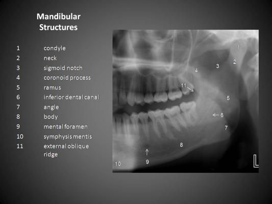
However complications may arise due to an extension anterior to the mental foramen that forms the mandible incisive canal MIC. Incisive foramen is the opening of the incisive canal located immediately behind the maxillary central incisors. Exit through Foramina of Stenson.
All of the above F. The incisive foramen also known as nasopalatine foramen or anterior palatine foramen is the oral opening of the nasopalatine canalIt is located in the maxilla in the incisive fossa midline in the palate posterior to the central incisors at the junction of the medial palatine and incisive sutures. The incisive foramen is important because it is a potential site of cyst formation.
Anterior palatine foramen or nasopalatine foramen is the opening of the incisive canals on the hard palate immediately behind the incisor teethIt gives passage blood vessels and nerves. A working knowledge of normal anatomy of the oral-facial region as it appears on radiographs is essential in assessing accurately the information. NASOPALATINE duct cysts are cysts which form in the incisor canal region of the maxilla and originate in the nasopalatine duct or its remnants.
None of the above. The terminal branches of the greater palatine arteries pass through the. Radiographic anatomy lecture Slide 3 Incisive foramen opening of incisive canal containing sphenopalatine artery nasopalatine nerve which gives sensation to the anterior palate-Incisive foramen is the dark spot on perio apical radiograph- it is radiolucent as it is a canal and less bone in region so less dense fewer x-rays are attenuated Slide 4 Canine fossa- area where the bone.
These cysts have no direct relationship to the teeth but in their growth may encroach upon the incisor apices. The region between mental foramens is considered as a zone of choice for implants. Article in Croatian Cvetković T.
Anterior nasal spine 16. Intraoral Radiographic Anatomy Steven R. This chapter presents the major landmarks commonly found on conventional dental x-ray images.
E incisive foramen f median palatal suture b a d c facial view palatal view e f Landmarks in the Maxilla Incisive foramen Median palatine suture Pterygoid plates Pterodactyl gr. This program is Normal Radiographic Anatomy of Maxillary Periapical Projections This unit presents an introductory identification of the normal anatomy seen in maxillary periapical radiographs. In some radiographsB the incisive canal Fig.
In radiographs exposed from the region of the cuspid or lateral incisor the incisive foramen may appear as a radiolucency at the apex of one of the incisors. The incisive foramen shown as two foramina by Hebel and Stromberg 1976 lies in the midline of the hard palate between the left and right premaxillae and just behind the upper incisor teeth. On a _____ periapical radiograph the incisive foramen appears as a small ovoid or round _____ area located between the roots of the central incisors.
The incisive nerve innervates the anterior palatal soft tissues. The mean width of bone anterior to the incisive canal was 632 143 mm. Singer DDS 2123055674 srs2columbiaedu.
Transmit nasopalatine nerves and branches of the descending palatine artery. Tap card to see definition. The incisive foramen is situated within the incisive fossa of the maxilla.

Normal Anatomy On The Panoramic Radiograph Dentaltown Anatomical Landmarks On A Panoramic Radiograph Odontologia Estomatologia Estudiar Odontologia

Radiography Panoramic Radiograph Panoramic

Dental Radiographic Landmarks Dental Registered Dental Hygienist Dental Hygiene Student

Resultat De Recherche D Images Pour Skull Ap X Ray Anatomy Radiologic Technology Nuclear Medicine Anatomy

Pin By Windy Rothmund On Dental Hygiene Butterfly Shape Sphenoid Bone Palatine


0 Response to "incisive foramen radiograph"
Post a Comment